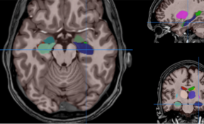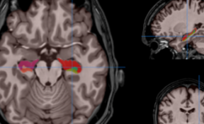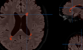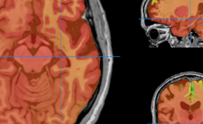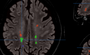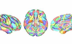
ASSEMBLYNET : Software providing segmentation of the intracranial cavity, brain, cortical lobes, cortical and subcortical structures using a T1w MR image
Domain Health and wellbeing
Technologies
Imaging Technologies for Health
Digital exploitation of health data
Challenges
Our software produces automatic quantitative analysis of MRI data for researchers and practitioners. This automatic and quantitative analysis of brain structure can be useful for many pathologies. Based on the latest advances in AI, AssemblyNet has a huge potential since is able to automatically extract all the brain structures in few minutes (12min). It has to be noted that AssemblyNet is one the most accurate available software. Its performance reaches human expert accuracy, and the produced segmentations are robust to pathology such as Alzheimer’s disease. The volume of brain structures has the potential to be an important biomarker for several neurological diseases (multiple sclerosis, Alzheimer’s disease, Parkinson, etc).
Innovative solution
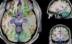
AssemblyNet is a pipeline of processes aimed to automatically analyze MRI brain data. It works as a black box from the user point of view as it gets an anonymized MRI brain volume in NIFTI format and produces a pdf report with the volumes of all the brain structures. The average processing time of the whole pipeline is around 12 minutes.
APPLICATIONS
-
Clinical trials
-
Companion diagnostics
-
Clinical routine
COMPETITIVES ADVANTAGES
-
No need of learning complex software packages
-
No need having expensive computational infrastructures in your local site
-
Faster and more accurate than other solutions
-
AssemblyNet works in a fully automatic manner and is able to provide brain structure volumes without any human interaction
-
Two reports (CSV and PDF) providing all the volumetry values calculated from the segmentations as well as asymmetry indexes
How it works
AssemblyNet provides full brain structure segmentation using T1w MR images thanks to a large ensemble of deep learning networks. AssemblyNet is shared as a docker image. The user provides a 3D T1, as a compressed nifti file, to the system. The system will first preprocess the image (denoising, inhomogeneity correction, registration) [using volbrain v1.0 preprocessing] and then will segment 133 brain structures.

In this protocol, the segmentation of subcortical structures follows the “general segmentation” protocol as defined by the MGH Center for Morphometric Analysis. Moreover, the segmentation of cortical structures follows the BrainCOLOR protocol. In AssemblyNet, the cortical structures are fused to produce the segmentation of cortex lobes. Finally, cortical and subcortical structures are fused to produce the segmentation of brain tissues.
AssemblyNet is also integrated within the web-service volBrain. There, the user uploads a 3D T1, as an anonymized nifti file, to the system. In the same way, the system preprocesses and segments the image. After few minutes, the results are sent back to the user by email.

Inventors
Developed by Pierrick COUPÉ (LaBRI - université de Bordeaux, CNRS, Bordeaux INP) & Jose Vicente MANJON HERRERA (Universitat Politècnica de València)
IP
Software registered with the Program Protection Agency APP
PARTERSHIPS
License to companies providing :
- Global healthcare solutions
- Diagnostic medical imaging solutions
- Medical imaging equipment
Contact
Jean-Luc CHAGNAUD
%6a%6c%2e%63%68%61%67%6e%61%75%64%40%61%73%74%2d%69%6e%6e%6f%76%61%74%69%6f%6e%73%2e%63%6f%6d
Associated innovations
- VolBrain A software to analyze MRI brain data. It provides a report including volume information on brain structures.
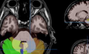
- CERES : A software to automatically analyze the cerebellum in brain MRI to provide a report including volume information on different aspects of the cerebellum
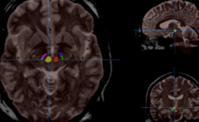
- pBrain : A software providing Parkinson’s disease related deep nucleus segmentation (Substantia Nigra locus niger), Red Nucleus and Subthalamic nucleus) using T2w MR images.
- HIPS : A software providing hippocampus subfield segmentation based on two different delineation protocols using both monomodal (T1w) and multimodal (T1w and T2w) MR images.
- LesionBrain : A software providing IC extraction, brain tissue classification and delineation/ classification of white matter hyperintense lesions using T1w and FLAIR MR images.
More…
AssemblyNet is available on : www.volbrain.net

